Single periapical radiographs are often made of individual teeth or groups of teeth to obtain information for treatment or diagnosis of localized diseases or abnormalities. Inclusion criteria included periapical X-ray images of permanents teeth and patients aged 14 years old with good sharpness.

How Make Periapical X Ray
Paralleling Technique for Periapical X-rays The paralleling technique results in good quality x-rays with a minimum of distortion and is the most reliable technique for taking periapical x-rays.

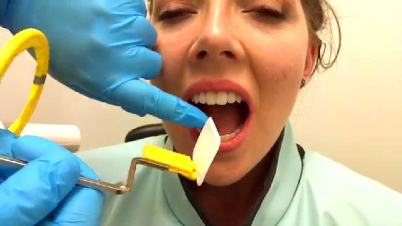
. Assessment of relationship of roots to various vital structures. Periapical X-rays are used to detect any abnormalities of the root structure and surrounding bone structure. To take a periapical exposure the hygienist or x-ray technician places a small photosensitive imaging plate coated with phosphorus into a sterile wrapper and inserts it into the patients mouth just like a conventional X-ray film card.
The film is placed parallel to the long axis of the tooth to be radiographed and the central beam of X-ray is directed at right angle to the film and the teeth. Changes to the angulation of the X-ray beam in relation to the teeth and film can help diagnosis and treatment by producing images which provide additional information not alway. In these situations the bisecting angle technique may be used.
The X-ray is taken and the exposed plate is then loaded into a scanner or processor which reads the image. Periapical views are used to record the crowns roots and surrounding bone. Periapical radiographs provide important information about the teeth and surrounding bone.
Students are given a demonstration of panoramic radiology. The patient was positioned upright with hisher mouth was opened as wide as possible to allow the X-ray beam to pass to the sensor unobstructed from the opposite side of the mouth. Most frequently used radiography is for the periapical which is performed by the bisecting Thus when considering the execution of the radiographic technique and the possibility of errors that occur during the exposure of X-ray image XR receptors it is important to identify those that occur more frequently.
Film parallel to the long axis of the teeth and guides the central ray of the x-ray beam to be directed at a right angle to the teeth and the receptor. The film is placed parallel to the long axis of the tooth in question and the central x-ray beam should be directed perpendicular to the long axis of the tooth. Assessment of root formation n completion.
These X-rays are used to find dental problems below the gum line or in the jaw such as impacted teeth tooth fractures abscesses tumours and bone changes linked to some diseases. Film development and mounting are discussed and practiced. The paralleling technique results in good quality x-rays with a minimum of distortion and is the most reliable technique for taking periapical x-rays.
Implant site assessment and. The bisecting short-cone and paralleling long-cone techniques are two of the most commonly used techniques. With this technique the film is placed parallel to the long axis of a tooth allowing the X-ray to be focused perpendicular to the long axis of the tooth.
The patient is seated upright in the dental chair and should remove any removable dental appliances glasses or jewelry that could interfere with the X-ray beam. Periapical radiographic techniques during endodontic diagnosis and treatment Int Endod J. Periapical X-rays.
For this purpose a special technique of periapical radiography was developed by Gordon M. Different techniques and instruments are used to drain and decompress large periapical lesions ranging from placing a stainless steel tube into the root canal exhibiting persistent apical exudation 202 204 which is non-surgical decompression to placing polyvinyl or polyethylene tubes through the alveolar mucosa covering the apical lesion which is surgical. Periapical X-rays show the entire tooth from the exposed crown to the end of the root and the bones that support the tooth.
By using a film sensor holder with still. Ensure they are seated high enough so it is easy to see the occlusal. Parallel technique The image receptor is placed in a holder and placed in the mouth parallel to the longitudinal axis of the tooth under.
Occlusal X-rays show full tooth development and placement 9. By using a filmsensor holder with fixed image receptor and. Exclusion criteria were periapical X-ray images of tooth germs or images which have distortion effects.
RADIOGRAPHS Periapical Bitewing Occlusal 2. The extraoral periapical radiographic technique was performed for both maxillary and mandibular teeth using Newman and Friedman technique2. The X-ray tubehead is then aimed at right angles vertically and horizontally to both the tooth and the image.
Extraoral radiograph Panoramic X-ray Tomograms Cephalometric projections Sialography Computed tomography 10. Periapical radiography is a commonly used intraoral imaging technique in radiology and may be a component of your radiologic examination. 2 Central x-ray entry is point down from the outer corner of the eye to the occlusal plane 3 Use size 2 media 4 Displays the distal of the second premolars to the distal of the last molars 5 Clearly shows the crowns and crestal bone 6 Contacts are seen clearly 7 Occlusal plane is parallel to film edge.
Periapical film is held parallel to the long axis of the tooth using film-holding instruments. Radiographic techniques 1. Assessment of root morphology.
Both techniques have advantages and disadvantages. Periapical images have been collected using the FONA X70 Intraoral X-rays machine and PSPIX Imaging Plates. The X-ray head is directed at right angles vertically and horizontally of both the tooth and the image receptor.
The image receptor is placed in a holder and positioned in the mouth parallel to the long axis of the tooth under. The Bisecting Angle Technique is an alternative to the paralleling technique for taking periapical films. The long cone paralleling technique positions the receptor ie.
The paralleling technique is recommended for routine periapical radiography but there are some instances when it is very difficult due to patient anatomy or lack of cooperation. The sensor was placed on the. The film is placed parallel to the long axis of the tooth in question and the central x-ray beam should be directed perpendicular to the long axis of the tooth.
Fitzgerald called as paralleling or long cone technique. This method produces images of the teeth on the receptor with minimal distortion. The paralleling technique results in good quality x-rays with a minimum of distortion and is the most reliable technique for taking periapical x-rays.
Students learn how to expose dental periapical x-ray film using the Rinn Paralleling Technique using the XCP film holder. Periapical views are used to record the crowns roots and surrounding bone.
How To Take Periapical Radiographs Youtube

Periapical Radiography Pocket Dentistry

Periapical Radiography Pocket Dentistry
Periapical Radiography Pocket Dentistry

Periapical Radiography Pocket Dentistry

Periapical Radiography Pocket Dentistry

Periapical Radiography Pocket Dentistry

Periapical Radiography Pocket Dentistry
0 comments
Post a Comment